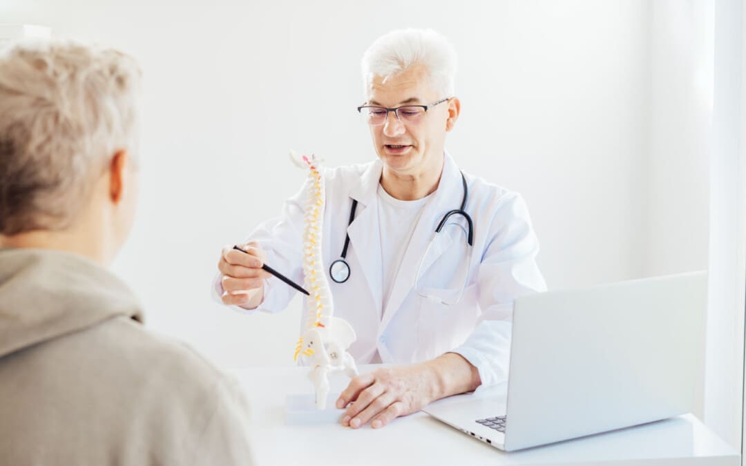
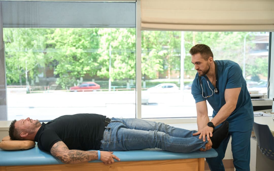
Back Clinic Chiropractic. Đây là một hình thức điều trị thay thế tập trung vào việc chẩn đoán và điều trị các chấn thương và tình trạng cơ xương khác nhau, đặc biệt là những bệnh liên quan đến cột sống. Tiến sĩ Alex Jimenez thảo luận về cách điều chỉnh cột sống và thao tác bằng tay thường xuyên có thể giúp cải thiện và loại bỏ nhiều triệu chứng có thể gây khó chịu cho cá nhân. Các bác sĩ nắn khớp xương tin rằng trong số những lý do chính gây ra đau và bệnh là do sự lệch trục của các đốt sống trong cột sống (đây được gọi là hiện tượng lệch cột sống).
Thông qua việc sử dụng phát hiện bằng tay (hoặc sờ nắn), áp dụng cẩn thận áp lực, xoa bóp và thao tác thủ công các đốt sống và khớp (được gọi là điều chỉnh), các chuyên gia nắn khớp xương có thể giảm bớt áp lực và kích thích lên dây thần kinh, phục hồi khả năng vận động của khớp và giúp trả lại cân bằng nội môi của cơ thể . Từ lệch cột sống, hoặc lệch cột sống, đến đau thần kinh tọa, một tập hợp các triệu chứng dọc theo dây thần kinh tọa do sự chèn ép dây thần kinh, chăm sóc chỉnh hình có thể dần dần khôi phục trạng thái tự nhiên của cá nhân. Tiến sĩ Jimenez biên soạn một nhóm các khái niệm về trị liệu thần kinh cột sống để giáo dục tốt nhất cho các cá nhân về nhiều loại chấn thương và tình trạng ảnh hưởng đến cơ thể con người.
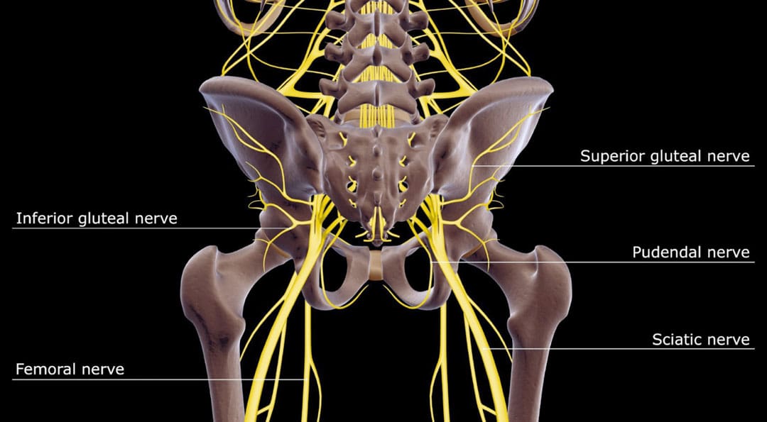
For individuals experiencing pelvic pain, it could be a disorder of the pudendal nerve known as pudendal neuropathy or neuralgia that leads to chronic pain. The condition can be caused by pudendal nerve entrapment, where the nerve becomes compressed or damaged. Can knowing the symptoms help healthcare providers correctly diagnose the condition and develop an effective treatment plan?
Pudendal Neuropathy
The pudendal nerve is the main nerve that serves the perineum, which is the area between the anus and the genitalia – the scrotum in men and the vulva in women. The pudendal nerve runs through the gluteus muscles/buttocks and into the perineum. It carries sensory information from the external genitalia and the skin around the anus and perineum and transmits motor/movement signals to various pelvic muscles. (Origoni, M. et al., 2014) Pudendal neuralgia, also referred to as pudendal neuropathy, is a disorder of the pudendal nerve that can lead to chronic pelvic pain.
Nguyên nhân
Chronic pelvic pain from pudendal neuropathy can be caused by any of the following (Kaur J. et al., 2024)
Excessive sitting on hard surfaces, chairs, bicycle seats, etc. Bicyclists tend to develop pudendal nerve entrapment.
Trauma to the buttocks or pelvis.
Sinh đẻ.
Bệnh thần kinh đái tháo đường.
Bony formations that push against the pudendal nerve.
Thickening of ligaments around the pudendal nerve.
Các triệu chứng
Pudendal nerve pain can be described as stabbing, cramping, burning, numbness, or pins and needles and can present (Kaur J. et al., 2024)
In the perineum.
In the anal region.
In men, pain in the scrotum or penis.
In women, pain in the labia or vulva.
Trong khi giao hợp.
When urinating.
During a bowel movement.
When sitting and goes away after standing up.
Because the symptoms are often hard to distinguish, pudendal neuropathy can often be hard to differentiate from other types of chronic pelvic pain.
Cyclist’s Syndrome
Prolonged sitting on a bicycle seat can cause pelvic nerve compression, which can lead to chronic pelvic pain. The frequency of pudendal neuropathy (chronic pelvic pain caused by entrapment or compression of the pudendal nerve) is often referred to as Cyclist’s Syndrome. Sitting on certain bicycle seats for long periods places significant pressure on the pudendal nerve. The pressure can cause swelling around the nerve, which causes pain and, over time, can lead to nerve trauma. Nerve compression and swelling can cause pain described as burning, stinging, or pins and needles. (Durante, J. A., and Macintyre, I. G. 2010) For individuals with pudendal neuropathy caused by bicycling, symptoms can appear after prolonged biking and sometimes months or years later.
Take breaks at least 20–30 seconds after each 20 minutes of riding.
While riding, change positions frequently.
Stand up to pedal periodically.
Take time off between riding sessions and races to rest and relax the pelvic nerves. 3–10 day breaks can help in recovery. (Durante, J. A., and Macintyre, I. G. 2010)
If pelvic pain symptoms are barely starting to develop, rest and see a healthcare provider or specialist for an examination.
Seat
Use a soft, wide seat with a short nose.
Have the seat level or tilted slightly forward.
Seats with cutout holes place more pressure on the perineum.
If numbness or pain is present, try a seat without holes.
Lắp xe đạp
Adjust the seat height so the knee is slightly bent at the bottom of the pedal stroke.
The body’s weight should rest on the sitting bones/ischial tuberosities.
Keeping the handlebar height below the seat can reduce pressure.
The Triathlon bike’s extreme-forward position should be avoided.
A more upright posture is better.
Mountain bikes have been associated with an increased risk of erectile dysfunction than road bikes.
Shorts
Wear padded bike shorts.
Phương pháp điều trị
A healthcare provider may use a combination of treatments.
The neuropathy can be treated with rest if the cause is excessive sitting or cycling.
Injury Medical Chiropractic and Functional Medicine Clinic care plans and clinical services are specialized and focused on injuries and the complete recovery process. Our areas of practice include Wellness and nutrition, Chronic Pain, Personal Injury, Auto Accident Care, Work Injuries, Back Injury, Low Back Pain, Neck Pain, Migraine Headaches, Sports Injuries, severe sciatica, Scoliosis, Complex Herniated Discs, Fibromyalgia, Chronic Pain, Complex Injuries, Stress Management, and Functional Medicine Treatments. If the individual requires other treatment, they will be referred to a clinic or physician best suited for their condition, as Dr. Jimenez has teamed with the top surgeons, clinical specialists, medical researchers, therapists, trainers, and premiere rehabilitation providers.
Mang thai và đau thần kinh tọa
dự án
Origoni, M., Leone Roberti Maggiore, U., Salvatore, S., & Candiani, M. (2014). Neurobiological mechanisms of pelvic pain. BioMed research international, 2014, 903848. doi.org/10.1155/2014/903848
Durante, J. A., & Macintyre, I. G. (2010). Pudendal nerve entrapment in an Ironman athlete: a case report. The Journal of the Canadian Chiropractic Association, 54(4), 276–281.
Chiaramonte, R., Pavone, P., & Vecchio, M. (2021). Diagnosis, Rehabilitation and Preventive Strategies for Pudendal Neuropathy in Cyclists, A Systematic Review. Journal of functional morphology and kinesiology, 6(2), 42. doi.org/10.3390/jfmk6020042
Đối với những người đã hết tất cả các lựa chọn điều trị khác cho chứng đau thắt lưng và chèn ép rễ thần kinh, liệu phẫu thuật cột sống bằng laser có thể giúp giảm bớt tình trạng chèn ép dây thần kinh và giúp giảm đau lâu dài không?
Phẫu thuật cột sống bằng Laser
Phẫu thuật cột sống bằng laser là một thủ thuật phẫu thuật xâm lấn tối thiểu, sử dụng tia laser để cắt xuyên qua và loại bỏ các cấu trúc cột sống đang chèn ép dây thần kinh và gây đau dữ dội. Thủ thuật xâm lấn tối thiểu thường ít gây đau đớn, tổn thương mô và phục hồi nhanh hơn so với các ca phẫu thuật mở rộng hơn.
Cách thức Hoạt động
Các thủ thuật xâm lấn tối thiểu giúp ít để lại sẹo và tổn thương các cấu trúc xung quanh, thường làm giảm các triệu chứng đau và thời gian hồi phục ngắn hơn. (Stern, J. 2009) Các vết mổ nhỏ được thực hiện để tiếp cận các cấu trúc cột sống. Với phẫu thuật hở lưng, một vết mổ lớn được thực hiện ở phía sau để tiếp cận cột sống. Phẫu thuật này khác với các phẫu thuật khác ở chỗ chùm tia laser, chứ không phải các dụng cụ phẫu thuật khác, được sử dụng để cắt các cấu trúc ở cột sống. Tuy nhiên, vết mổ ban đầu xuyên qua da được thực hiện bằng dao mổ. Laser là từ viết tắt của Khuếch đại ánh sáng được kích thích bằng sự phát xạ. Tia laser có thể tạo ra nhiệt độ cao để cắt xuyên qua các mô mềm, đặc biệt là những mô có hàm lượng nước cao, như đĩa đệm cột sống. (Stern, J. 2009) Đối với nhiều ca phẫu thuật cột sống, tia laser không thể được sử dụng để cắt xuyên xương vì nó tạo ra tia lửa tức thời có thể làm hỏng các cấu trúc xung quanh. Thay vào đó, phẫu thuật cột sống bằng laser chủ yếu được sử dụng để thực hiện phẫu thuật cắt bỏ đĩa đệm, đây là một kỹ thuật phẫu thuật nhằm loại bỏ một phần đĩa đệm bị phồng hoặc thoát vị đang đẩy vào các rễ thần kinh xung quanh, gây chèn ép dây thần kinh và đau thần kinh tọa. (Stern, J. 2009)
Rủi ro phẫu thuật
Phẫu thuật cột sống bằng laser có thể giúp giải quyết nguyên nhân gây chèn ép rễ thần kinh, nhưng làm tăng nguy cơ tổn thương các cấu trúc gần đó. Rủi ro liên quan bao gồm: (Brouwer, PA và cộng sự, 2015)
Nhiễm trùng
Chảy máu
Các cục máu đông
Các triệu chứng còn lại
Triệu chứng quay trở lại
Tổn thương thần kinh thêm
Tổn thương màng xung quanh tủy sống.
Cần phẫu thuật bổ sung
Chùm tia laser không chính xác như các dụng cụ phẫu thuật khác và đòi hỏi phải thực hành thành thạo và kiểm soát để tránh tổn thương tủy sống và rễ thần kinh. (Stern, J. 2009) Vì tia laser không thể cắt xuyên qua xương nên các dụng cụ phẫu thuật khác thường được sử dụng quanh các góc và ở các góc khác nhau vì chúng hiệu quả hơn và cho phép độ chính xác cao hơn. (Não và cột sống Đại Tây Dương, 2022)
Mục đích
Phẫu thuật cột sống bằng laser được thực hiện để loại bỏ các cấu trúc gây chèn ép rễ thần kinh. Nén rễ thần kinh có liên quan đến các tình trạng sau (Phòng khám Cleveland. 2018)
Đĩa phồng
Phình nang
đau thần kinh tọa
Hẹp ống sống
Khối u tủy sống
Rễ thần kinh bị tổn thương hoặc bị tổn thương và liên tục gửi tín hiệu đau mãn tính có thể được cắt bỏ bằng phẫu thuật laser, được gọi là cắt bỏ dây thần kinh. Tia laser đốt cháy và phá hủy các sợi thần kinh. (Stern, J. 2009) Vì phẫu thuật cột sống bằng laser bị hạn chế trong việc điều trị một số rối loạn cột sống nên hầu hết các thủ thuật cột sống xâm lấn tối thiểu đều không sử dụng tia laser. (Não và cột sống Đại Tây Dương. 2022)
Chuẩn bị
Đội ngũ phẫu thuật sẽ cung cấp hướng dẫn chi tiết hơn về những việc cần làm trong những ngày và giờ trước khi phẫu thuật. Để thúc đẩy quá trình lành thương tối ưu và phục hồi suôn sẻ, bệnh nhân nên vận động, ăn uống lành mạnh và ngừng hút thuốc trước khi phẫu thuật. Các cá nhân có thể cần ngừng dùng một số loại thuốc để ngăn ngừa chảy máu quá nhiều hoặc tương tác với thuốc mê trong quá trình phẫu thuật. Thông báo cho nhà cung cấp dịch vụ chăm sóc sức khỏe về tất cả các đơn thuốc, thuốc không kê đơn và thuốc bổ sung đang được sử dụng.
Phẫu thuật cột sống bằng laser là một thủ tục ngoại trú tại bệnh viện hoặc trung tâm phẫu thuật ngoại trú. Bệnh nhân có thể sẽ về nhà vào cùng ngày phẫu thuật. (Phòng khám Cleveland. 2018) Bệnh nhân không thể lái xe đến hoặc rời khỏi bệnh viện trước hoặc sau khi phẫu thuật, vì vậy hãy sắp xếp để gia đình hoặc bạn bè đưa đón. Giảm thiểu căng thẳng và ưu tiên sức khỏe tinh thần và cảm xúc lành mạnh là điều quan trọng để giảm viêm và hỗ trợ phục hồi. Bệnh nhân càng khỏe mạnh thì phẫu thuật càng dễ dàng hồi phục và phục hồi chức năng.
Mong đợi
Cuộc phẫu thuật sẽ do bệnh nhân và nhà cung cấp dịch vụ chăm sóc sức khỏe quyết định và lên lịch tại bệnh viện hoặc trung tâm phẫu thuật ngoại trú. Sắp xếp cho một người bạn hoặc thành viên gia đình lái xe đến nơi phẫu thuật và về nhà.
Trước khi phẫu thuật
Bệnh nhân sẽ được đưa đến phòng tiền phẫu và được yêu cầu thay áo choàng.
Bệnh nhân sẽ được khám sức khỏe ngắn gọn và trả lời các câu hỏi về bệnh sử.
Bệnh nhân nằm trên giường bệnh và y tá đặt ống truyền tĩnh mạch để truyền thuốc và chất lỏng.
Kíp phẫu thuật sẽ sử dụng giường bệnh để vận chuyển bệnh nhân ra vào phòng mổ.
Đội ngũ phẫu thuật sẽ hỗ trợ bệnh nhân lên bàn mổ và bệnh nhân sẽ được gây mê.
Bệnh nhân có thể nhận được gây mê toàn thân, sẽ khiến bệnh nhân ngủ để phẫu thuật, hoặc gây tê vùng, tiêm vào cột sống để làm tê vùng bị ảnh hưởng. (Phòng khám Cleveland. 2018)
Đội ngũ phẫu thuật sẽ khử trùng vùng da nơi thực hiện vết mổ.
Dung dịch sát trùng sẽ được sử dụng để diệt vi khuẩn và ngăn ngừa nguy cơ nhiễm trùng.
Sau khi vệ sinh, cơ thể sẽ được phủ khăn tiệt trùng để giữ cho vết mổ luôn sạch sẽ.
Trong khi phẫu thuật
Để phẫu thuật cắt bỏ đĩa đệm, bác sĩ phẫu thuật sẽ rạch một vết nhỏ dài dưới một inch bằng dao mổ dọc theo cột sống để tiếp cận các rễ thần kinh.
Một dụng cụ phẫu thuật gọi là nội soi là một camera được đưa vào vết mổ để quan sát cột sống. (Brouwer, PA và cộng sự, 2015)
Sau khi xác định được phần đĩa có vấn đề gây ra tình trạng nén, tia laser sẽ được đưa vào để cắt xuyên qua phần đó.
Phần đĩa cắt được lấy ra và vị trí vết mổ được khâu lại.
Sau Phẫu thuật
Sau phẫu thuật, bệnh nhân được đưa đến phòng hồi sức, nơi các dấu hiệu sinh tồn được theo dõi khi thuốc mê hết tác dụng.
Sau khi ổn định, bệnh nhân thường có thể về nhà một hoặc hai giờ sau khi phẫu thuật.
Bác sĩ phẫu thuật sẽ xác định khi nào cá nhân đó có thể tiếp tục lái xe.
Phục hồi
Sau khi phẫu thuật cắt bỏ đĩa đệm, cá nhân có thể trở lại làm việc trong vòng vài ngày đến vài tuần, tùy thuộc vào mức độ nghiêm trọng, nhưng có thể mất đến ba tháng để trở lại hoạt động bình thường. Thời gian hồi phục có thể dao động từ hai đến bốn tuần hoặc ít hơn nếu tiếp tục công việc ít vận động hoặc từ 12 đến XNUMX tuần đối với công việc đòi hỏi thể chất nhiều hơn và phải nâng vật nặng. (Trường Y và Y tế Công cộng thuộc Đại học Wisconsin, 2021) Trong hai tuần đầu tiên, bệnh nhân sẽ được đưa ra những hạn chế để tạo điều kiện cho cột sống phục hồi cho đến khi ổn định hơn. Các hạn chế có thể bao gồm: (Trường Y và Y tế Công cộng thuộc Đại học Wisconsin, 2021)
Không uốn, xoắn hoặc nâng.
Không hoạt động thể chất vất vả, bao gồm tập thể dục, làm việc nhà, làm vườn và quan hệ tình dục.
Không uống rượu trong giai đoạn đầu hồi phục hoặc trong khi dùng thuốc giảm đau có chất gây nghiện.
Không lái xe hoặc vận hành phương tiện cơ giới cho đến khi thảo luận với bác sĩ phẫu thuật.
Nhà cung cấp dịch vụ chăm sóc sức khỏe có thể đề nghị vật lý trị liệu để thư giãn, tăng cường và duy trì sức khỏe cơ xương. Vật lý trị liệu có thể thực hiện hai đến ba lần mỗi tuần trong bốn đến sáu tuần.
Quy trình xét duyệt
Các khuyến nghị phục hồi tối ưu bao gồm:
Ngủ đủ giấc, ít nhất bảy đến tám giờ.
Duy trì thái độ tích cực và học cách đối phó và quản lý căng thẳng.
Duy trì độ ẩm cho cơ thể.
Thực hiện theo chương trình tập luyện theo chỉ định của bác sĩ vật lý trị liệu.
Thực hành các tư thế lành mạnh bằng cách ngồi, đứng, đi và ngủ.
Duy trì hoạt động và hạn chế thời gian ngồi. Cố gắng đứng dậy và đi bộ 1-2 giờ một lần trong ngày để duy trì hoạt động và ngăn ngừa cục máu đông. Tăng dần thời gian hoặc khoảng cách khi quá trình phục hồi diễn ra.
Đừng thúc ép phải làm quá nhiều việc quá sớm. Việc gắng sức quá mức có thể làm tăng cơn đau và làm chậm quá trình phục hồi.
Học các kỹ thuật nâng đúng cách để sử dụng cơ lõi và cơ chân nhằm ngăn ngừa áp lực tăng lên cột sống.
Thảo luận về các lựa chọn điều trị để kiểm soát các triệu chứng với nhà cung cấp dịch vụ chăm sóc sức khỏe hoặc chuyên gia để xác định xem phẫu thuật cột sống bằng laser có phù hợp hay không. Chấn thương y tế Các kế hoạch chăm sóc và dịch vụ lâm sàng của Phòng khám Y tế Chỉnh hình và Y học Chức năng đều chuyên biệt và tập trung vào các chấn thương cũng như quá trình phục hồi hoàn toàn. Tiến sĩ Jimenez đã hợp tác với các bác sĩ phẫu thuật hàng đầu, chuyên gia lâm sàng, nhà nghiên cứu y tế, nhà trị liệu, huấn luyện viên và nhà cung cấp dịch vụ phục hồi chức năng hàng đầu. Chúng tôi tập trung vào việc khôi phục các chức năng bình thường của cơ thể sau chấn thương và tổn thương mô mềm bằng cách sử dụng các Phương pháp Chiropractic Chuyên biệt, Chương trình Chăm sóc Sức khỏe, Dinh dưỡng Chức năng và Tích hợp, Huấn luyện Thể dục Nhanh nhẹn và Vận động cũng như Hệ thống Phục hồi chức năng cho mọi lứa tuổi. Các lĩnh vực hành nghề của chúng tôi bao gồm Sức khỏe & Dinh dưỡng, Đau mãn tính, Chấn thương cá nhân, Chăm sóc tai nạn ô tô, Chấn thương khi làm việc, Chấn thương lưng, Đau thắt lưng, Đau cổ, Đau nửa đầu, Chấn thương khi chơi thể thao, Đau thần kinh tọa nặng, Vẹo cột sống, Thoát vị đĩa đệm phức tạp, Đau cơ xơ hóa, Mãn tính Đau đớn, Chấn thương phức tạp, Kiểm soát căng thẳng, Điều trị bằng thuốc chức năng và các phác đồ chăm sóc trong phạm vi.
Brouwer, PA, Brand, R., van den Akker-van Marle, ME, Jacobs, WC, Schenk, B., van den Berg-Huijsmans, AA, Koes, BW, van Buchem, MA, Arts, MP, & Peul , WC (2015). Giải nén đĩa đệm bằng laser qua da so với phẫu thuật cắt bỏ vi phẫu thông thường ở bệnh đau thần kinh tọa: một thử nghiệm ngẫu nhiên có đối chứng. Tạp chí cột sống: tạp chí chính thức của Hiệp hội cột sống Bắc Mỹ, 15(5), 857–865. doi.org/10.1016/j.spinee.2015.01.020
Trường Y và Y tế Công cộng thuộc Đại học Wisconsin. (2021). Hướng dẫn chăm sóc tại nhà sau phẫu thuật cắt bỏ phần thắt lưng, giải nén hoặc cắt bỏ đĩa đệm. bệnh nhân.uwhealth.org/healthfacts/4466
Individuals may discover a lump, bump, or nodule under the skin around their lower back, hips, and sacrum that can cause pain by compressing nerves and damaging the fascia. Can knowing the conditions linked to them and their symptoms help healthcare providers determine a correct diagnosis and develop an effective treatment plan for experiencing?
Painful Bumps, Nodules Around Low Back, Hips, and Sacrum
Painful masses in and around the hips, the xương mông, and the lower back are lumps of fat or lipomas, fibrous tissue, or other types of nodules that move when pressed on. Some healthcare providers and chiropractors, in particular, use the non-medical term back mice (In 1937, the term was used to describe lumps associated with episacroiliac lipoma) to describe the bumps. Some healthcare professionals argue against calling the masses mice because it is not specific and could lead to misdiagnoses or incorrect treatment.
Most show up in the lower back and hip area.
In some cases, they protrude or herniate through the lumbodorsal fascia or the network of connective tissue that covers the deep muscles of the lower and middle back.
Other lumps can develop in the tissue under the skin.
Today, many conditions are associated with back mice lumps, including:
Iliac crest pain syndrome
Multifidus triangle syndrome
Lumbar fascial fat herniation
Lumbosacral (sacrum) fat herniation
Episacral lipoma
Các điều kiện liên quan
Iliac Crest Pain Syndrome
Also known as iliolumbar syndrome, iliac crest pain syndrome develops when a tear in the ligament occurs.
The ligament band connects the fourth and fifth lumbar vertebrae with the ilium on the same side. (Dąbrowski, K. Ciszek, B. 2023)
Nguyên nhân bao gồm:
Tearing the ligament from repeated bending and twisting.
Trauma or fracture of the ilium bone caused by a fall or vehicle collision accident.
Multifidus Triangle Syndrome
Multifidus triangle syndrome develops when the multifidus muscles along the spine weaken and diminish function or ability.
These muscles can atrophy, and intramuscular fatty tissue can replace the muscle.
The lumbodorsal fascia is a thin fibrous membrane covering the back’s deep muscles.
Lumbar fascial fat herniation is a painful mass of fat that protrudes or herniates through the membrane, gets trapped and inflamed, and causes pain.
The causes of this type of herniation are currently unknown.
Lumbosacral (Sacrum) Fat Herniation
Lumbosacral describes where the lumbar spine meets the sacrum.
Lumbosacral fat herniation is a painful mass like lumbar facial herniation in a different location around the sacrum.
The causes of this type of herniation are currently unknown.
Episacral Lipoma
Episacral lipoma is a small painful nodule under the skin that primarily develops over the top outer edges of the pelvic bone. These lumps occur when a portion of the dorsal fat pad protrudes through a tear in the thoracodorsal fascia, the connective tissue that helps hold the back muscles in place. (Erdem, H. R. et al., 2013) A healthcare provider may refer an individual to an orthopedist or orthopedic surgeon for this lipoma. An individual may also find pain relief from a massage therapist familiar with the condition. (Erdem, H. R. et al., 2013)
Các triệu chứng
Back lumps can often be seen under the skin. They are typically tender to the touch and can make sitting in a chair or lying on the back difficult, as they often appear on the hip bones and sacroiliac region. (Bicket, M. C. et al., 2016) The nodules may:
Be firm or tight.
Have an elastic feel.
Move under the skin when pressed.
Cause intense, severe pain.
The pain results from pressure on the lump, which compresses the nerves.
Damage to the underlying fascia can also cause pain symptoms.
Chẩn đoán
Some individuals do not realize they have nodules or lumps until pressure is applied. Chiropractors and massage therapists often find them during treatments but do not diagnose the abnormal fatty growth. The chiropractor or massage therapist will refer the patient to a qualified dermatologist or medical professional who can perform imaging studies and a biopsy. Determining what the lumps are can be challenging because they are non-specific. Healthcare providers sometimes diagnose the nodules by injecting them with a local anesthetic. (Bicket, M. C. et al., 2016)
Chẩn đoán phân biệt
The fatty deposits can be any number of things, and the same applies to the sources of nerve pain. A healthcare provider may further diagnose by ruling out other causes, which can include:
Sebaceous Cysts
A benign, fluid-filled capsule between the layers of skin.
Subcutaneous Abscess
A collection of pus beneath the skin.
Usually painful.
It can become inflamed.
đau thần kinh tọa
Radiating nerve pain down one or both legs that is caused by a herniated disc, bone spur, or spasming muscles in the lower back.
ung thư mỡ
Malignant tumors can sometimes appear as fatty growths in the muscles.
Liposarcoma is typically diagnosed by biopsy, where some tissue is removed from the nodule and examined for cancer cells. (Johns Hopkins Medicine. 2024)
An MRI or CT scan may also be performed to determine the exact location of the nodule.
Painful lipomas are also associated with fibromyalgia.
Điều trị
Back nodules are usually benign, so there’s no reason to remove them unless they’re causing pain or mobility problems (American Academy of Orthopedic Surgeons: OrthoInfo. 2023). However, they should be examined to make sure they are not cancerous. Treatment usually involves injected anesthetics, such as lidocaine or corticosteroids, as well as over-the-counter pain relievers like NSAIDs.
Phẫu thuật
If pain is severe, surgical removal may be recommended. This involves cutting out the mass and repairing the fascia for lasting relief. However, removal may not be recommended if there are many nodules, as some individuals can have hundreds. Liposuction may be effective if the lumps are smaller, more extensive, and comprise more fluid. (American Family Physician. 2002) Complications of surgical removal can include:
Sẹo
Bầm tím
Kết cấu da không đồng đều
Nhiễm trùng
Complementary and Alternative Treatment
Complimentary and Alternative Medicine treatments like acupuncture, dry needling, and spinal manipulation can help. Many chiropractors believe back nodules can be successfully treated with complementary and alternative therapies. A common approach uses acupuncture and spinal manipulation in combination. A case study reported that anesthetic injections followed by dry needling, which is similar to acupuncture, improved pain relief. (Bicket, M. C. et al., 2016)
Injury Medical Chiropractic and Functional Medicine Clinic specializes in progressive therapies and functional rehabilitation procedures focused on restoring normal body functions after trauma and soft tissue injuries and the complete recovery process. Our areas of practice include Wellness & Nutrition, Chronic Pain, Personal Injury, Auto Accident Care, Work Injuries, Back Injury, Low Back Pain, Neck Pain, Migraine Headaches, Sports Injuries, Severe Sciatica, Scoliosis, Complex Herniated Discs, Fibromyalgia, Chronic Pain, Complex Injuries, Stress Management, Functional Medicine Treatments, and in-scope care protocols. If the individual requires other treatment, they will be referred to a clinic or physician best suited for their condition, as Dr. Jimenez has teamed with the top surgeons, clinical specialists, medical researchers, therapists, trainers, and premiere rehabilitation providers.
Ngoài bề mặt
dự án
Dąbrowski, K., & Ciszek, B. (2023). Anatomy and morphology of iliolumbar ligament. Surgical and radiologic anatomy : SRA, 45(2), 169–173. doi.org/10.1007/s00276-022-03070-y
Seyedhoseinpoor, T., Taghipour, M., Dadgoo, M., Sanjari, M. A., Takamjani, I. E., Kazemnejad, A., Khoshamooz, Y., & Hides, J. (2022). Alteration of lumbar muscle morphology and composition in relation to low back pain: a systematic review and meta-analysis. The spine journal : official journal of the North American Spine Society, 22(4), 660–676. doi.org/10.1016/j.spinee.2021.10.018
Erdem, H. R., Nacır, B., Özeri, Z., & Karagöz, A. (2013). Episakral lipoma: Bel ağrısının tedavi edilebilir bir nedeni [Episacral lipoma: a treatable cause of low back pain]. Agri : Agri (Algoloji) Dernegi’nin Yayin organidir = The journal of the Turkish Society of Algology, 25(2), 83–86. doi.org/10.5505/agri.2013.63626
Bicket, M. C., Simmons, C., & Zheng, Y. (2016). The Best-Laid Plans of “Back Mice” and Men: A Case Report and Literature Review of Episacroiliac Lipoma. Pain physician, 19(3), 181–188.
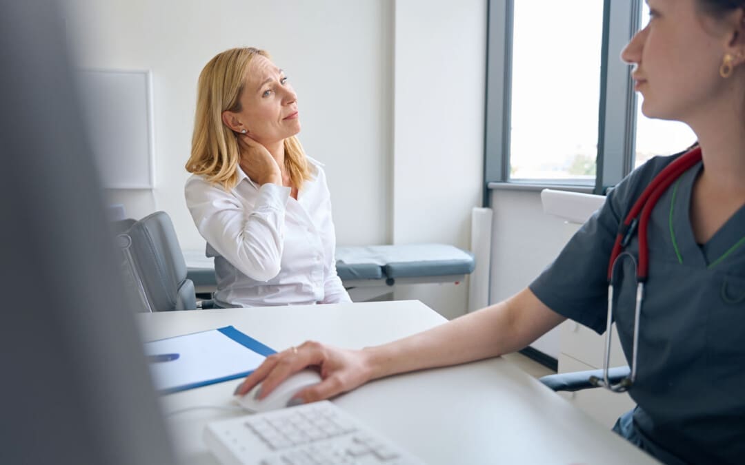
Khi bị đau thần kinh tọa hoặc các cơn đau thần kinh lan tỏa khác, việc học cách phân biệt giữa đau dây thần kinh và các loại đau khác nhau có thể giúp cá nhân nhận biết khi nào rễ thần kinh cột sống bị kích thích hoặc bị nén hoặc các vấn đề nghiêm trọng hơn cần được chăm sóc y tế?
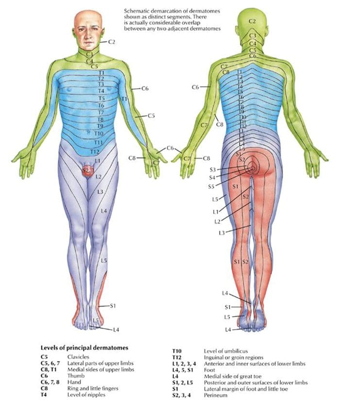
Rễ thần kinh cột sống và da liễu
Các tình trạng cột sống như thoát vị đĩa đệm và hẹp ống sống có thể dẫn đến cơn đau lan xuống một cánh tay hoặc chân. Các triệu chứng khác bao gồm yếu, tê và/hoặc cảm giác như bị điện giật hoặc nóng rát. Thuật ngữ y học cho các triệu chứng dây thần kinh bị chèn ép là bệnh rễ thần kinh (Viện Y tế Quốc gia: Viện Rối loạn Thần kinh và Đột quỵ Quốc gia. 2020). Dermatomes có thể góp phần gây kích ứng ở tủy sống, nơi rễ thần kinh gây ra các triệu chứng ở lưng và tay chân.
Giải Phẫu
Tủy sống có 31 đoạn.
Mỗi đoạn có rễ thần kinh ở bên phải và bên trái cung cấp các chức năng vận động và cảm giác cho các chi.
Các nhánh thông trước và sau kết hợp với nhau tạo thành các dây thần kinh cột sống thoát ra khỏi ống đốt sống.
31 đoạn cột sống tạo ra 31 dây thần kinh cột sống.
Mỗi cơ truyền đầu vào dây thần kinh cảm giác từ một vùng da cụ thể ở bên đó và vùng cơ thể.
Những vùng này được gọi là da liễu.
Ngoại trừ dây thần kinh cột sống cổ thứ nhất, mỗi dây thần kinh cột sống đều có các tế bào da liễu.
Các dây thần kinh cột sống và các lớp da liên quan của chúng tạo thành một mạng lưới khắp cơ thể.
Mục Đích của Dermatomes
Dermatomes là vùng cơ thể/da với đầu vào cảm giác được chỉ định cho từng dây thần kinh cột sống. Mỗi rễ thần kinh có một tế bào da liên quan và các nhánh khác nhau cung cấp năng lượng cho từng tế bào da từ rễ thần kinh đó. Dermatomes là con đường qua đó thông tin giật gân trong da truyền tín hiệu đến và từ hệ thống thần kinh trung ương. Những cảm giác được cảm nhận về mặt vật lý, như áp suất và nhiệt độ, sẽ được truyền đến hệ thần kinh trung ương. Khi rễ thần kinh cột sống bị nén hoặc bị kích thích, thường là do nó tiếp xúc với một cấu trúc khác, sẽ dẫn đến bệnh lý rễ thần kinh. (Viện Y tế Quốc gia: Viện Rối loạn Thần kinh và Đột quỵ Quốc gia. 2020).
Bệnh lý thần kinh
Bệnh rễ thần kinh mô tả các triệu chứng do dây thần kinh bị chèn ép dọc theo cột sống. Các triệu chứng và cảm giác phụ thuộc vào vị trí dây thần kinh bị chèn ép và mức độ chèn ép.
Cổ tử cung
Đây là hội chứng đau và/hoặc suy giảm cảm giác vận động khi rễ thần kinh ở cổ bị nén.
Nó thường biểu hiện bằng cơn đau lan xuống một cánh tay.
Các cá nhân cũng có thể trải qua các cảm giác điện như kim châm, sốc và cảm giác nóng rát, cũng như các triệu chứng vận động như yếu và tê.
Ngang lưng
Bệnh rễ thần kinh này là kết quả của sự chèn ép, viêm hoặc tổn thương dây thần kinh cột sống ở lưng dưới.
Cảm giác đau, tê, ngứa ran, cảm giác điện hoặc nóng rát và các triệu chứng vận động như yếu di chuyển xuống một chân là phổ biến.
Chẩn đoán
Một phần của khám thực thể bệnh lý rễ thần kinh là kiểm tra cảm giác ở da. Người học viên sẽ sử dụng các bài kiểm tra thủ công cụ thể để xác định mức độ cột sống mà các triệu chứng bắt nguồn. Khám thủ công thường đi kèm với các xét nghiệm chẩn đoán hình ảnh như MRI, có thể cho thấy những bất thường ở rễ thần kinh cột sống. Khám sức khoẻ toàn diện sẽ xác định xem rễ thần kinh cột sống có phải là nguồn gốc của các triệu chứng hay không.
Điều trị các nguyên nhân cơ bản
Nhiều chứng rối loạn ở lưng có thể được điều trị bằng các liệu pháp bảo tồn để giúp giảm đau hiệu quả. Ví dụ, đối với trường hợp thoát vị đĩa đệm, người bệnh có thể được khuyên nên nghỉ ngơi và dùng thuốc chống viêm không steroid. Châm cứu, vật lý trị liệu, nắn khớp xương, kéo không phẫu thuật, hoặc liệu pháp giải nén cũng có thể được quy định. Đối với những cơn đau dữ dội, các cá nhân có thể được tiêm steroid ngoài màng cứng để giảm đau bằng cách giảm viêm. (Học viện phẫu thuật chỉnh hình Hoa Kỳ: OrthoInfo. 2022) Đối với chứng hẹp cột sống, trước tiên nhà cung cấp có thể tập trung vào vật lý trị liệu để cải thiện thể lực tổng thể, tăng cường cơ bụng và cơ lưng cũng như duy trì chuyển động ở cột sống. Thuốc giảm đau, bao gồm NSAID và thuốc tiêm corticosteroid, có thể làm giảm viêm và giảm đau. (Đại học Thấp khớp Hoa Kỳ. 2023) Các nhà vật lý trị liệu cung cấp nhiều liệu pháp khác nhau để giảm các triệu chứng, bao gồm giải nén và lực kéo bằng tay và cơ học. Phẫu thuật có thể được khuyến nghị cho những trường hợp bệnh lý rễ thần kinh không đáp ứng với các phương pháp điều trị bảo tồn.
Chấn thương y tế Các kế hoạch chăm sóc và dịch vụ lâm sàng của Phòng khám Y tế Chỉnh hình và Y học Chức năng đều chuyên biệt và tập trung vào các chấn thương cũng như quá trình phục hồi hoàn toàn. Các lĩnh vực hành nghề của chúng tôi bao gồm Sức khỏe & Dinh dưỡng, Đau mãn tính, Chấn thương cá nhân, Chăm sóc tai nạn ô tô, Chấn thương khi làm việc, Chấn thương lưng, Đau thắt lưng, Đau cổ, Đau nửa đầu, Chấn thương khi chơi thể thao, Đau thần kinh tọa nặng, Vẹo cột sống, Thoát vị đĩa đệm phức tạp, Đau cơ xơ hóa, Mãn tính Đau đớn, Chấn thương phức tạp, Kiểm soát căng thẳng, Điều trị bằng thuốc chức năng và các phác đồ chăm sóc trong phạm vi. Chúng tôi tập trung vào việc khôi phục các chức năng bình thường của cơ thể sau chấn thương và tổn thương mô mềm bằng cách sử dụng các Phương pháp Chiropractic Chuyên biệt, Chương trình Chăm sóc Sức khỏe, Dinh dưỡng Chức năng và Tích hợp, Huấn luyện Thể dục và Phục hồi Chức năng cho mọi lứa tuổi. Nếu cá nhân đó cần phương pháp điều trị khác, họ sẽ được giới thiệu đến một phòng khám hoặc bác sĩ phù hợp nhất với tình trạng của họ. Tiến sĩ Jimenez đã hợp tác với các bác sĩ phẫu thuật, chuyên gia lâm sàng, nhà nghiên cứu y tế, nhà trị liệu, huấn luyện viên và nhà cung cấp dịch vụ phục hồi chức năng hàng đầu để mang El Paso, phương pháp điều trị lâm sàng hàng đầu đến cộng đồng của chúng ta.
Đòi lại khả năng vận động của bạn: Chăm sóc chỉnh hình để phục hồi đau thần kinh tọa
For individuals who suffer from migraine headaches, can incorporating physical therapy help decrease pain, improve mobility, and manage future attacks?
Migraine Physical Therapy
Cervicogenic migraine headaches can cause pain, limited motion, or confusing symptoms like dizziness or nausea. They may originate from the neck or cervical spine and be called cervicogenic headaches. A chiropractic physical therapy team can assess the spine and offer treatments that help improve mobility and decrease pain. Individuals may benefit from working with a migraine physical therapy team to perform treatments for specific conditions, quickly and safely relieving pain and returning to their previous level of activity.
Cervical Spine Anatomy
The neck is comprised of seven stacked cervical vertebrae. The cervical vertebrae protect the spinal cord and allow the neck to move through:
Flexion
Extension
Rotation
Uốn bên
The upper cervical vertebrae help support the skull. There are joints on either side of the cervical level. One connects to the back of the skull and allows motion. This suboccipital area is home to several muscles that support and move the head, with nerves that travel from the neck through the suboccipital area into the head. The nerves and muscles in this area may be a source of neck pain and/or headaches.
Các triệu chứng
Sudden motions can trigger symptoms of cervicogenic migraine, or they may come on during sustained neck postures. (Page P. 2011) The symptoms are often dull and non-throbbing and may last several hours to days. Symptoms of cervicogenic migraine headache may include:
Pain on both sides of the back of the head.
Pain in the back of the head that radiates to one shoulder.
Pain on one side of the upper neck that radiates to the temple, forehead, or eye.
Pain in one side of the face or cheek.
Reduced range of motion in the neck.
Độ nhạy sáng hoặc âm thanh
Buồn nôn
Chóng mặt hoặc chóng mặt
Chẩn đoán
Tools a physician may use may include:
X-quang
MRI
Chụp CT
Physical examination includes neck range of motion and palpation of the neck and skull.
When first visiting a physical therapist, they will go through medical history and conditions, and questions will be asked about the onset of pain, symptom behavior, medications, and diagnostic studies. The therapist will also ask about previous treatments and review medical and surgical history. Components of the evaluation may include:
Palpation of the neck and skull
Measures of neck range of motion
Strength measurements
Postural assessment
Once the evaluation is completed, the therapist will work with the individual to develop a personalized treatment program and rehabilitation goals. Various treatments are available.
Tập thể dục
Exercises to improve neck motion and decrease pressure on cervical nerves may be prescribed and may include. (Park, S. K. et al., 2017)
Xoay cổ tử cung
Cervical flexion
Cervical side bending
Cervical retraction
The therapist will train the individual to move slowly and steadily and avoid sudden or jerky movements.
Postural Correction
If forward head posture is present, the upper cervical spine and the suboccipital area could compress the nerves that travel up the back of the skull. Correcting posture may be an effective strategy for treatment and can include:
Performing targeted postural exercises.
Utilizing a supportive neck pillow for sleep.
Using a lumbar support when sitting.
Kinesiology taping may help increase tactile awareness of back and neck position and improve overall postural awareness.
Nhiệt / Băng
Heat or ice may be applied to the neck and skull to help decrease pain and inflammation.
Heat can help relax tight muscles and improve circulation and may be used before performing neck stretches.
xoa bóp
If tight muscles are limiting neck motion and causing head pain, a massage can help improve mobility.
A special technique called suboccipital release loosens the muscles that attach the skull to the neck for improved motion and decreased nerve irritation.
Manual and Mechanical Traction
Part of the migraine physical therapy plan may involve mechanical or manual traction to decompress the neck’s discs and joints, improve motion in the neck, and decrease pain.
Joint mobilizations may be used to improve neck motion and manage pain. (Paquin, J. P. 2021)
Kích thích điện
Electrical stimulation, like electro-acupuncture or transcutaneous neuromuscular electrical stimulation, may be used on the neck muscles to decrease pain and improve headache symptoms.
Therapy Duration
Most migraine physical therapy sessions for cervicogenic headaches last about four to six weeks. Individuals may experience relief within a few days of starting therapy, or symptoms may come and go in different phases for weeks. Some experience continued migraine headache pain for months after starting treatment and use techniques they learned to help control symptoms.
Injury Medical Chiropractic and Functional Medicine Clinic specializes in progressive therapies and functional rehabilitation procedures focused on restoring normal body functions after trauma and soft tissue injuries. We use Specialized Chiropractic Protocols, Wellness Programs, Functional and integrative Nutrition, Agility and mobility Fitness Training, and Rehabilitation Systems for all ages. Our natural programs use the body’s ability to achieve specific measured goals. We have teamed up with the city’s premier doctors, therapists, and trainers to provide high-quality treatments that empower our patients to maintain the healthiest way of living and live a functional life with more energy, a positive attitude, better sleep, and less pain.
Chăm sóc chỉnh hình cho chứng đau nửa đầu
dự án
Trang P. (2011). Đau đầu cổ tử cung: một cách tiếp cận dựa trên bằng chứng để quản lý lâm sàng. Tạp chí quốc tế về vật lý trị liệu thể thao, 6(3), 254–266.
Headache Classification Committee of the International Headache Society (IHS) (2013). The International Classification of Headache Disorders, 3rd edition (beta version). Cephalalgia : an international journal of headache, 33(9), 629–808. doi.org/10.1177/0333102413485658
Rana M. V. (2013). Managing and treating headache of cervicogenic origin. The Medical clinics of North America, 97(2), 267–280. doi.org/10.1016/j.mcna.2012.11.003
Park, S. K., Yang, D. J., Kim, J. H., Kang, D. H., Park, S. H., & Yoon, J. H. (2017). Effects of cervical stretching and cranio-cervical flexion exercises on cervical muscle characteristics and posture of patients with cervicogenic headache. Journal of physical therapy science, 29(10), 1836–1840. doi.org/10.1589/jpts.29.1836
Paquin, J. P., Tousignant-Laflamme, Y., & Dumas, J. P. (2021). Effects of SNAG mobilization combined with a self-SNAG home-exercise for the treatment of cervicogenic headache: a pilot study. The Journal of manual & manipulative therapy, 29(4), 244–254. doi.org/10.1080/10669817.2020.1864960
Footwear can cause lower back pain and problems for some individuals. Can understanding the connection between footwear and back problems help individuals find the right shoes to maintain back health and relieve pain?
Footwear Back Pain
The back provides the strength for physical activities. Back pain affects daily life and can have various causes. Unhealthy posture, walking, twisting, turning, bending, and reaching can contribute to back problems that result in pain. According to the CDC, 39% of adults report living with back pain (Trung tâm kiểm soát và phòng ngừa dịch bệnh, 2019). Improper footwear can also contribute to back pain. Selecting footwear carefully can help bring pain relief and help maintain spinal health. Individuals can enjoy less pain and manage symptoms by choosing shoes that maintain spinal alignment and protect the feet from blunt impact.
Understanding the Back Pain-Footwear Connection
Improper footwear could be the cause of lower back pain. What impacts the bones at the bottom of the neuromusculoskeletal system radiates upward and affects the spine and back muscles. What footwear is used travels upward, impacting gait, posture, spinal alignment, and more. When back problems originate from the feet, these are biomechanical issues. Biomechanics means how the bones, joints, and muscles work together and how changes in external forces impact the body.
Phong trào
When the feet impact the ground, they are the first extremities to absorb shock for the rest of the body. Individuals will start to walk differently if they have a problem or change in their feet. Wearing shoes with improper support can increase the wear and tear on the muscles and joints, leading to awkward and unnatural movement. For example, consider the difference between standing on tiptoes in high heels and the natural flat-footed state. Well-cushioned shoes help absorb impact and lessen pain sensations. The pressures on each of the joints shift balance, which causes instability problems with less pressure on some and more on others. This creates an imbalance that leads to pain and joint conditions.
Tư thế
Maintaining a healthy posture is another factor in preventing or alleviating back pain. With the right footwear, the body can maintain a healthier stance and the right curvature throughout the spine, and it helps distribute the weight evenly. This results in decreased stress on ligaments, muscles, and joints. (Nhà xuất bản Y tế Harvard. 2014) It’s recommended to see an orthopedist to get to the root of an individual’s condition. For some, a herniated disc, sciatica, automobile collision, fall, unhealthy ergonomics, or a combination, as well as other underlying issues, may be contributing to their back pain.
Shoe Types and Their Impact on The Back
How various shoes impact posture, potentially causing or relieving back pain.
Cao gót
High heels can definitely contribute to back pain. They change body posture, causing a domino effect on the spine. The body’s weight is shifted to increase pressure on the balls of the feet, and the spine’s alignment becomes altered. High heels also affect how the ankles, knees, and hips move when walking, balance, and how the back muscles operate, all of which can worsen back pain.
Giày phẳng
Flat shoes may not be the best choice for spinal health. If they lack arch support, they can cause the foot to roll inward, known as pronation. This can contribute to misalignment, which can strain the knees, hips, and lower back. However, they can be a decent choice if they provide arch support. When wearing flat shoes with healthy support, the weight is distributed evenly on the feet and the spine. This helps maintain correct posture, which can help prevent and/or alleviate back pain.
Sneakers, Tennis, and Athletic Shoes
Sneakers, tennis, and giày thể thao can relieve back pain with thorough cushioning and support. Choosing the right ones involves determining the activity that will be done in them. There are tennis, running, basketball, pickleball, skating shoes, and more. Research what features will be needed for the sport or activity. This could include:
Heel cups
Insole cushioning
Cơ sở rộng
Other features to meet individual foot needs.
It is recommended that athletic shoes be changed every 300 to 500 miles of walking or running or with any signs of unevenness when placed on a flat surface, as worn-out soles and degraded materials can increase the risk of injury and back pain. (American Academy of Podiatric Sports Medicine, 2024). If a certain pair puts the legs, hips, or ankles into an unnatural position or impedes regular movement, it may be time to replace them.
Chọn giày phù hợp
The ideal solution for choosing shoe wear is to get a gait analysis and a review of how you walk and run. Various healthcare professionals may offer this service to tailor each individual’s search for the right shoes for back pain. In gait analysis, individuals are asked to run and walk, sometimes on camera, while a professional notes physical tendencies, like when the foot hits the ground and whether it rolls inward or outward. This provides data on affected posture, movement, pain levels, how much arch support is needed, and what type to wear to help prevent back pain. Once the analysis is complete, it will guide you on what to look for, such as what level of arch support, heel height, or material is best for you.
Injury Medical Chiropractic and Functional Medicine Clinic specializes in progressive, cutting-edge therapies and functional rehabilitation procedures focused on clinical physiology, total health, practical strength training, and complete conditioning. We focus on restoring normal body functions after trauma and soft tissue injuries. We use Specialized Chiropractic Protocols, Wellness Programs, Functional and integrative Nutrition, Agility and mobility Fitness Training, and Rehabilitation Systems for all ages. Our programs are natural and use the body’s ability to achieve specific measured goals rather than introducing harmful chemicals, controversial hormone replacement, unwanted surgeries, or addictive drugs. We have teamed up with the city’s premier doctors, therapists, and trainers to provide high-quality treatments that empower our patients to maintain the healthiest way of living and live a functional life with more energy, a positive attitude, better sleep, and less pain.
Lợi ích của việc sử dụng phương pháp chỉnh hình bàn chân tùy chỉnh
For individuals who are dealing with back pain and problems, could knowing how to improve and maintain intervertebral disc health help alleviate symptoms?
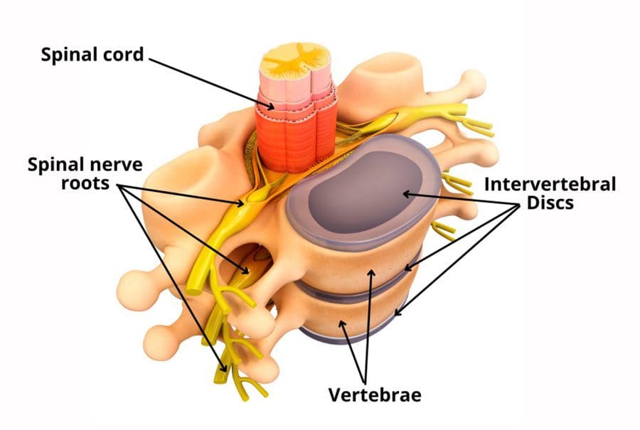
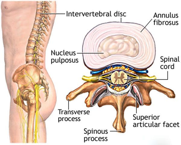
Intervertebral Disc Health
The spinal column comprises 24 movable bones and 33 bones called vertebrae. The vertebral bones are stacked on top of each other. The intervertebral disc is the cushioning substance between the adjacent bones. (Dartmouth. 2008)
Bones
The vertebral bones are small and round in an area called the vertebral body. In the back is a bony ring from which protrusions extend and arches and pathways are formed. Each structure has one or more purposes and includes: (Waxenbaum JA, Reddy V, Williams C, et al., 2023)
Stabilizing the spine.
Providing a space for the connective tissue and back muscles to attach.
Providing a tunnel for the spinal cord to pass through cleanly.
Providing a space where nerves exit and branch out to all areas of the body.
Structure
The intervertebral disc is the cushioning that sits between the vertebrae. The design of the spine allows it to move in various directions:
Flexion or bending
Extension or arching
Tilting and rotation or twisting.
Powerful forces act upon and influence the spinal column to produce these movements. The intervertebral disc absorbs shock during movement and protects the vertebrae and spinal cord from injury and/or trauma.
Khả năng
On the outside, strong woven fiber tissues form an area called the annulus fibrosis. The annulus fibrosis contains and protects the softer gel substance in the center, the nucleus pulposus. (Y.S. Nosikova et al., 2012) The nucleus pulposis provides shock absorption, flexibility, and pliability, especially under pressure during spinal movement.
cơ học
The nucleus pulposus is a soft gel substance located in the center of the disc that allows elasticity and flexibility under stress forces to absorb compression. (Nedresky D, Reddy V, Singh G. 2024) The swivel action alters the tilt and rotation of the vertebra above and below, buffering the effects of spinal motion. The discs swivel in response to the direction the spine moves. The nucleus pulposus is made mostly of water, which moves in and out through small pores, acting as byways between the vertebra and disc bone. Body positions that load the spine, like sitting and standing, push the water out of the disc. Lying down on the back or in a supine position facilitates water restoration into the disc. As the body ages, the discs lose water/khử nước, leading to disc degeneration. The intervertebral disc has no blood supply, which means that for a disc to receive necessary nutrition and for waste removal, it must rely on water circulation to stay healthy.
Quan tâm
Some ways of maintaining intervertebral disc health include:
Paying attention to posture.
Changing positions frequently throughout the day.
Exercising and moving around.
Applying correct body mechanics to physical activities.
Sleeping on a supportive mattress.
Uống nhiều nước.
Ăn uống lành mạnh.
Duy trì cân nặng hợp lý.
Drinking alcohol in moderation.
Bỏ hút thuốc.
At Injury Medical Chiropractic and Functional Medicine Clinic, we treat injuries and chronic pain syndromes by improving an individual’s ability through flexibility, mobility, and agility programs tailored for all age groups and disabilities. Our chiropractic team, care plans, and clinical services are specialized and focused on injuries and the complete recovery process. Our areas of practice include Wellness & Nutrition, Acupuncture, Chronic Pain, Personal Injury, Auto Accident Care, Work Injuries, Back Injury, Low Back Pain, Neck Pain, Migraine Headaches, Sports Injuries, Severe Sciatica, Scoliosis, Complex Herniated Discs, Fibromyalgia, Chronic Pain, Complex Injuries, Stress Management, Functional Medicine Treatments, and in-scope care protocols. If other treatment is needed, individuals will be referred to a clinic or physician best suited to their injury, condition, and/or ailment.
Beyond the Surface: Understanding the Effects of Personal Injury
Waxenbaum, J. A., Reddy, V., Williams, C., & Futterman, B. (2024). Anatomy, Back, Lumbar Vertebrae. In StatPearls. www.ncbi.nlm.nih.gov/pubmed/29083618
Nosikova, Y. S., Santerre, J. P., Grynpas, M., Gibson, G., & Kandel, R. A. (2012). Characterization of the annulus fibrosus-vertebral body interface: identification of new structural features. Journal of anatomy, 221(6), 577–589. doi.org/10.1111/j.1469-7580.2012.01537.x
Công cụ Tìm Bác sĩ của IFM là mạng lưới giới thiệu lớn nhất trong Y học Chức năng, được tạo ra để giúp bệnh nhân xác định vị trí các bác sĩ Y học Chức năng ở bất kỳ đâu trên thế giới. Các bác sĩ được chứng nhận IFM được liệt kê đầu tiên trong kết quả tìm kiếm, nhờ trình độ học vấn sâu rộng của họ về Y học chức năng


 Shoe Types and Their Impact on The Back
 Khả năng


